
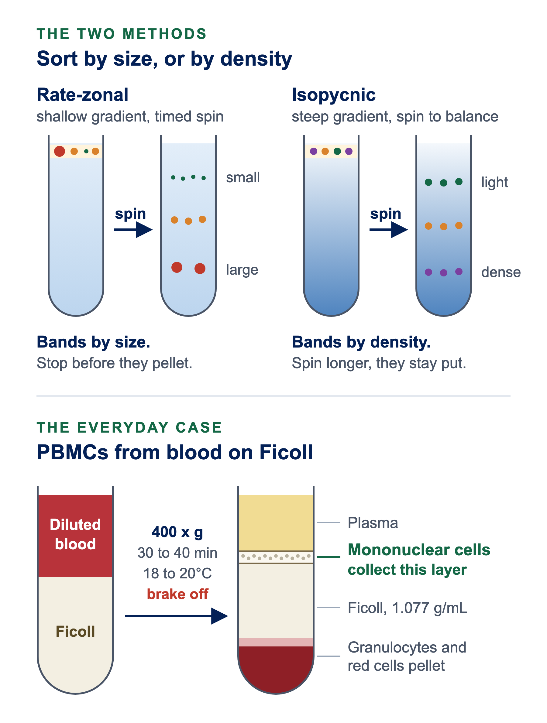
Density gradient centrifugation spins a sample through liquid that gets denser toward the bottom of the tube, so particles stop in separate bands. Rate-zonal sorts them by size; isopycnic sorts them by density.
Density gradient centrifugation separates cells, organelles, viruses and molecules by spinning them through a liquid that gets denser toward the bottom of the tube. Instead of everything piling into one pellet, particles stop partway down in bands you can collect. This guide covers the two ways to run it, how to choose a gradient medium, the Ficoll protocol most labs use to isolate mononuclear cells from blood, and how to fix a separation that goes wrong.
Key Facts
- Two methods: rate-zonal sorts particles by size, isopycnic sorts them by density
- Most common use: isolating peripheral blood mononuclear cells (PBMCs) on a 1.077 g/mL medium such as Ficoll-Paque
- Ficoll spin: 400 x g for 30 to 40 minutes at 18 to 20°C, brake off
- What to expect: about 60% of the mononuclear cells recovered, at around 95% purity and over 90% viability
- Common media: Ficoll, Percoll, sucrose, iodixanol and cesium chloride
01 · Theory
How density gradient centrifugation works
In an ordinary spin, everything denser than the liquid ends up in the pellet, and particles that differ only slightly come down together. A gradient changes that. The liquid in the tube is lightest at the top and densest at the bottom, so particles can be held apart in separate bands. How they are held apart depends on which of two methods you use.
Rate-zonal: sorting by size
The sample goes on top of a shallow gradient as a thin band. During the spin, large particles move down faster than small ones. The gradient is chosen so the particles are denser than the liquid at every point, which means nothing ever comes to rest: you stop the run before the separated zones reach the bottom of the tube.1 The gradient’s job here is to keep the moving bands from mixing, which is why a shallow sucrose gradient, typically 5 to 20%, is the classic choice.2
That makes rate-zonal a timed method. Stop too early and the zones have not separated. Run too long and they all reach the bottom together.
Isopycnic: sorting by density
Isopycnic means “equal density.” Here the gradient covers the full range of densities in your sample. Each particle moves until it reaches the point where the liquid’s density matches its own, and stops there. Particles are separated only by density, whatever their size.1 Because they stop, spinning longer does not push them into the pellet once they have arrived.
| Rate-zonal | Isopycnic | |
|---|---|---|
| Separates by | Size and shape (how fast particles sediment) | Buoyant density |
| Gradient | Shallow; lighter than every particle | Steep enough to span every particle’s density |
| When to stop | On time, before the zones reach the bottom | Once the bands have formed; extra time does no harm |
| Typical jobs | Cell components and macromolecules that differ in size2 | Cells on Percoll, DNA on cesium chloride, viruses on iodixanol1, 2, 3 |
Where the Ficoll method fits. The PBMC protocol below uses a single layer of medium rather than a full gradient. Mononuclear cells and platelets are not dense enough to enter a 1.077 g/mL layer, so they collect at its surface. Red blood cells are clumped together by the Ficoll and sink through it, and granulocytes, made denser by the slightly hypertonic medium, follow them.4 The principle is the same as isopycnic: cells are sorted by whether they are lighter or heavier than the medium.
The saltiness of the medium, its osmolality, changes the very thing you are measuring. In a concentrated medium, cells and other membrane-bound particles lose water and shrink. In a dilute one, they swell. Either way their apparent density shifts. Particles band at much lower densities in isotonic Percoll than in sucrose.1 That is why living cells are separated on media that can be kept at physiological osmolality, such as Ficoll-Paque or isotonic Percoll.
02 · Media
Choosing a gradient medium
The medium decides what you can separate, so choose it by your sample first and your budget second.
- Ficoll
- Blood cells. Ficoll-Paque PLUS is Ficoll PM400 mixed with sodium diatrizoate at a density of 1.077 g/mL, made for isolating mononuclear cells from human blood.5 A 1.084 g/mL version is also available.4for PBMCs
- Percoll
- Cells and organelles. Coated colloidal silica with very low osmolality, so it can be made isotonic throughout. It is non-toxic and covers densities from 1.0 to 1.3 g/mL. Spun at over 10,000 x g in a fixed-angle rotor, it forms its own gradient. Percoll PLUS is a newer version with a silane coating.1
- Sucrose
- Rate-zonal work on macromolecules and viruses. Readily available, with well-known properties, which makes it the standard for rate-zonal separations.6 The method itself began on sucrose in 1951, purifying a plant virus.7 Concentrated sucrose is hyperosmotic, so membrane-bound particles shrink in it.1
- Iodixanol
- Viruses. An iodinated medium, sold as OptiPrep, that stays iso-osmotic up to a density of 1.32 g/mL and has very low toxicity.8 A widely used method purifies AAV vectors on steps of 15, 25, 40 and 60% iodixanol, collecting the virus from the 40% step.3
- CsCl
- DNA. Cesium chloride forms a steep gradient sensitive enough to separate DNA labelled with a heavy isotope from unlabelled DNA.2 It is how Meselson and Stahl separated heavy and light DNA in 1958, spinning at 44,770 rpm for 20 hours.9
Making Percoll isotonic
Percoll is supplied in water, so it has to be made isotonic before cells touch it. Mix 9 parts Percoll with 1 part 1.5 M NaCl or 10x concentrated culture medium to make a stock isotonic Percoll, then dilute it to the density you need. For subcellular particles that clump in salt, use 1 part 2.5 M sucrose instead of the NaCl. Cytiva recommends checking the osmolality with an osmometer, and the final density with a densitometer or refractometer.1
03 · Protocol
Ficoll protocol: isolating PBMCs from blood, step by step
This is the method Cytiva gives for Ficoll-Paque PLUS. It isolates the lymphocytes and monocytes that most immunology work starts from.4
Run everything at 18 to 20°C. Too cold, and the red cells clump less and the medium is denser, so red cells and granulocytes stay in your cell layer. Too warm, and the medium is less dense, so some lymphocytes sink into it and viability can suffer.4 A medium taken straight from the refrigerator is too cold.
- 1.077
- Density. Ficoll-Paque PLUS, 1.077 ± 0.001 g/mL at 20°C.4
- 1:1
- Dilution. Blood mixed with an equal volume of balanced salt solution.4
- 400 x g
- Spin. 30 to 40 minutes at 18 to 20°C, in a swing-out rotor with the brake off.4
- 3 vol
- Wash. At least three volumes of balanced salt solution per volume of cells.4
What you need
- Blood treated with an anticoagulant, or defibrinated4
- A balanced salt solution such as HBSS, for diluting the blood and washing the cells
- Ficoll-Paque PLUS, warmed to 18 to 20°C
- A centrifuge with a swing-out rotor4
- Sterile 10 mL centrifuge tubes and pipettes
- Dilute the bloodIn a 10 mL tube, mix 2 mL of blood with 2 mL of balanced salt solution.4
- Add the FicollPut 3 mL of Ficoll-Paque in a fresh centrifuge tube.4
- Layer the blood on topSlowly run the 4 mL of diluted blood onto the Ficoll so the two do not mix.4 Holding the pipette tip against the tube wall, just above the liquid, keeps the boundary sharp.
- SpinCentrifuge at 400 x g for 30 to 40 minutes at 18 to 20°C, with the brake turned off.4
- Remove the plasmaDraw off the top layer of plasma and platelets with a sterile pipette, leaving the cloudy band of mononuclear cells undisturbed.4
- Collect the cellsTransfer the mononuclear cell band to a sterile centrifuge tube.4
- First washAdd at least three volumes of balanced salt solution (about 6 mL), mix gently with a pipette, and spin at 400 to 500 x g for 10 to 15 minutes at 18 to 20°C. Discard the liquid.4
- Second washResuspend the cells in 6 to 8 mL of balanced salt solution and spin for 10 minutes at 18 to 20°C. Use 400 to 500 x g for the best recovery, or 60 to 100 x g if you need to leave the platelets behind.4
Diluting blood or washing PBMCs?
Browse all buffers and salt solutions
Made in the USA and manufactured in house. Certificate of Analysis with every lot. Bulk pricing, purchase orders and custom formulations welcome.
Scaling up. Larger blood volumes work if you keep roughly the same layer heights: about 2.4 cm of Ficoll-Paque and 3.0 cm of diluted blood.4
What a good run gives you. Cytiva’s specification is 60 ± 20% of the mononuclear cells in the original blood, 95 ± 5% of them mononuclear, over 90% viable, with at most 5% granulocytes and 10% red cells.4
Watch it done
A short demonstration of the same method, from layering the blood to collecting the cell band at the interface.
Running other gradients
Percoll, sucrose and iodixanol separations follow the same outline, with a few choices the Ficoll method makes for you.
- Build the gradientFor a step gradient, layer solutions of decreasing density on top of each other, with a pipette or a syringe fitted with a wide-bore needle.1 For Percoll you can skip this: spin it in a fixed-angle rotor, for example at 30,000 x g for 15 minutes, and it forms a smooth gradient on its own.1
- Check the densitiesMeasure the densest and lightest solutions before you start. For Percoll, refractive index rises in a straight line with density, so a refractometer reading tells you the density.1
- Load the sampleFor rate-zonal runs, load it as a thin band on top.1 On a preformed Percoll gradient, cells band isopycnically in 15 to 20 minutes at 400 x g.1
- Pick the rotorUse a swinging-bucket rotor for rate-zonal runs that need the sharpest bands; fixed-angle and vertical rotors suit isopycnic work such as banding plasmid DNA in cesium chloride. In those, the gradient tips back to horizontal as the rotor stops, so decelerate slowly.6 Never try to form a Percoll gradient in a swinging bucket: at over 10,000 x g the silica simply pellets.1
- Collect the fractionsPipette bands off the top, puncture the bottom of the tube and collect the drops in order, or pump a dense solution under the gradient and take fractions off the top.1 To take one band from the middle, puncture the side of the tube just below it with a needle and syringe and draw it off.6
04 · Troubleshooting
Fuzzy bands, low yield and contamination: what went wrong
Most failed separations trace back to temperature, dilution or the rotor. Find your symptom below.
- Smeared
- Fuzzy or smeared bands. Something mixed the layers. In fixed-angle and vertical rotors the gradient tips back to horizontal as the rotor stops, so a hard stop smears it: decelerate slowly.6 In a rate-zonal run, the gradient is what keeps moving bands from mixing, so check that it was poured and not disturbed.2
- Impure
- Red cells or granulocytes in your PBMC band. The run was too cold. The medium was denser and the red cells clumped less, so they could not get through. Warm everything to 18 to 20°C.4
- Yield
- Few cells at the interface. Either the run was too warm, so lymphocytes sank into the Ficoll,4 or the blood was not diluted 1:1. Undiluted or very concentrated blood traps lymphocytes in the clumps of red cells.4
- PLT
- Platelets in the final preparation. They collect at the interface with the mononuclear cells. Do the second wash at 60 to 100 x g, which leaves them in the liquid.4
- Pellet
- Everything ended up at the bottom in a rate-zonal run. The run went on too long. In rate-zonal separations the particles never stop, so the run must end before the zones reach the bottom.1
- Flat
- Percoll pelleted instead of forming a gradient. It was spun at high speed in a swinging-bucket rotor. Use a fixed-angle rotor.1
- Density
- Bands sit at an unexpected density. Check the osmolality. In a concentrated medium, membrane-bound particles shrink, and in a dilute one they swell, which shifts where they band.1 Cells band at much lower densities in isotonic Percoll than in sucrose.1
Sources
- Cytiva. Cell separation media: Methodology and applications (Percoll handbook, CY14836). 2021.
- Alberts B, Johnson A, Lewis J, Raff M, Roberts K, Walter P. Molecular Biology of the Cell. 4th ed. Garland Science; 2002. Fractionation of Cells.
- Zolotukhin S, Byrne BJ, Mason E, et al. Recombinant adeno-associated virus purification using novel methods improves infectious titer and yield. Gene Ther. 1999;6(6):973-985. doi:10.1038/sj.gt.3300938
- Cytiva. Isolation of mononuclear cells: Methodology and applications (CY13988). 2025.
- Cytiva. Ficoll-Paque PLUS Cell Preparation (data file CY13571). 2020.
- Beckman Coulter. Rotors and Tubes for Beckman Coulter Preparative Ultracentrifuges (LR-IM-22). 2003.
- Brakke MK. Density gradient centrifugation: a new separation technique. J Am Chem Soc. 1951;73(4):1847-1848. doi:10.1021/ja01148a508. Context from Scholthof KBG, Jackson AO, Van Etten JL. Myron Kendall Brakke 1921-2007: A Biographical Memoir. National Academy of Sciences; 2011.
- Ford T, Graham J, Rickwood D. Iodixanol: a nonionic iso-osmotic centrifugation medium for the formation of self-generated gradients. Anal Biochem. 1994;220(2):360-366. doi:10.1006/abio.1994.1350
- Meselson M, Stahl FW. The replication of DNA in Escherichia coli. Proc Natl Acad Sci U S A. 1958;44(7):671-682. doi:10.1073/pnas.44.7.671