
The EcoRI restriction enzyme is one of those tools you can’t imagine modern molecular biology without. Think of it as a pair of molecular scissors, but with a critical difference: instead of cutting randomly, it cuts DNA with surgical precision at a very specific sequence. This ability to make clean, predictable cuts is what turned the abstract idea of genetic engineering into a daily reality in the lab.
Unlocking Genetic Engineering with EcoRI
Before tools like the EcoRI restriction enzyme, trying to manipulate DNA was like attempting to edit a single sentence in a book with a sledgehammer. You might be able to smash the page, but you had no control. EcoRI gave scientists the scalpel they needed.
It works by recognizing a unique six-base-pair sequence—its "address"—on a strand of DNA. Its power lies in finding that one specific address out of millions or billions of base pairs and cutting it perfectly every single time. This was the breakthrough that allowed scientists to reliably isolate a gene from one organism and paste it into another.
The Discovery That Launched an Industry
The story of EcoRI starts in a University of California, San Francisco lab back in 1970. A PhD student, Robert Yoshimori, was studying how Escherichia coli bacteria defend themselves against invading viruses. He managed to isolate an enzyme from a particular strain, RY13, that could chop up foreign DNA.
This enzyme was given the name EcoRI: 'E' for Escherichia, 'co' for coli, 'R' for the RY13 strain, and 'I' because it was the first restriction enzyme isolated from that strain. It was a landmark discovery, giving researchers one of their very first tools for targeted DNA cutting. You can explore more about this landmark discovery and its scientific context.
But the real magic wasn't just that it could cut DNA—it was how it cut. EcoRI creates what we now call "sticky ends."
Sticky ends are short, single-stranded overhangs of DNA left after the enzyme makes its cut. These ends are complementary, acting like molecular Velcro that makes it incredibly easy to ligate, or "paste," two different pieces of DNA together, as long as both were cut by EcoRI.
This simple feature became the engine for the entire field of recombinant DNA technology. It gave scientists a reliable workflow for building custom DNA molecules, paving the way for breakthroughs in:
- Disease Research: Creating models to study gene function by inserting or deleting specific genes.
- Therapeutic Development: Engineering bacteria to produce human proteins like insulin.
- Biomanufacturing: Programming cells to produce complex biologics like monoclonal antibodies.
Decades later, the EcoRI restriction enzyme is anything but a historical footnote. It remains a workhorse in labs everywhere, essential for routine cloning, plasmid construction, and complex genome editing workflows.
EcoRI at a Glance
For a quick reference, here’s a summary of the key properties of the EcoRI enzyme.
| Characteristic | Description |
|---|---|
| Source Organism | Escherichia coli R-strain (RY13) |
| Recognition Sequence | 5'-G↓AATTC-3' |
| Cleavage Pattern | Creates a 4-base 5' overhang ("sticky end") with the sequence AATT. |
| Reaction Temperature | 37°C |
| Heat Inactivation | 65°C for 20 minutes. |
| Supplied Buffer | Typically supplied with a 10X buffer like NEB's rCutSmart™ Buffer. |
| Star Activity | Can occur with high glycerol, high enzyme concentration, or non-optimal buffer conditions. |
This table captures the core information you need to have on hand when planning your experiments, from setting up the reaction to troubleshooting potential issues.
How EcoRI Recognizes and Cuts DNA
The real power of the EcoRI restriction enzyme comes down to its incredible specificity. Think of it like a molecular key that fits only one very specific lock in a genome billions of base pairs long. EcoRI tirelessly scans the DNA, ignoring everything until it finds its one and only target sequence.
That target is the six-base-pair sequence 5'-G'AATTC-3'. What’s special about this sequence is that it’s a palindrome—it reads the same forwards on one strand as it does backward on the complementary strand (3'-CTTAA'G-5'). This symmetry is the secret handshake that allows the enzyme, which works as a two-part protein called a dimer, to bind perfectly to both strands of the DNA double helix.
Once it's locked on, the enzyme makes its signature move. It doesn't just randomly break the DNA. Instead, it makes a precise, staggered cut between the Guanine (G) and Adenine (A) bases on each strand.
Creating the Famous Sticky Ends
This staggered cut is what produces the iconic "sticky ends" that have made the EcoRI restriction enzyme a lab staple for decades. After the cut, each new end of the DNA has a four-base, single-stranded overhang with the sequence 5'-AATT-3'.
These overhangs act like perfectly shaped Velcro strips. An AATT overhang from one piece of DNA will only naturally pair up with another AATT overhang. This complementary nature is the bedrock of recombinant DNA technology, allowing you to control exactly how different DNA fragments join together.
This concept map breaks down how the enzyme's origin, target, and function all connect.
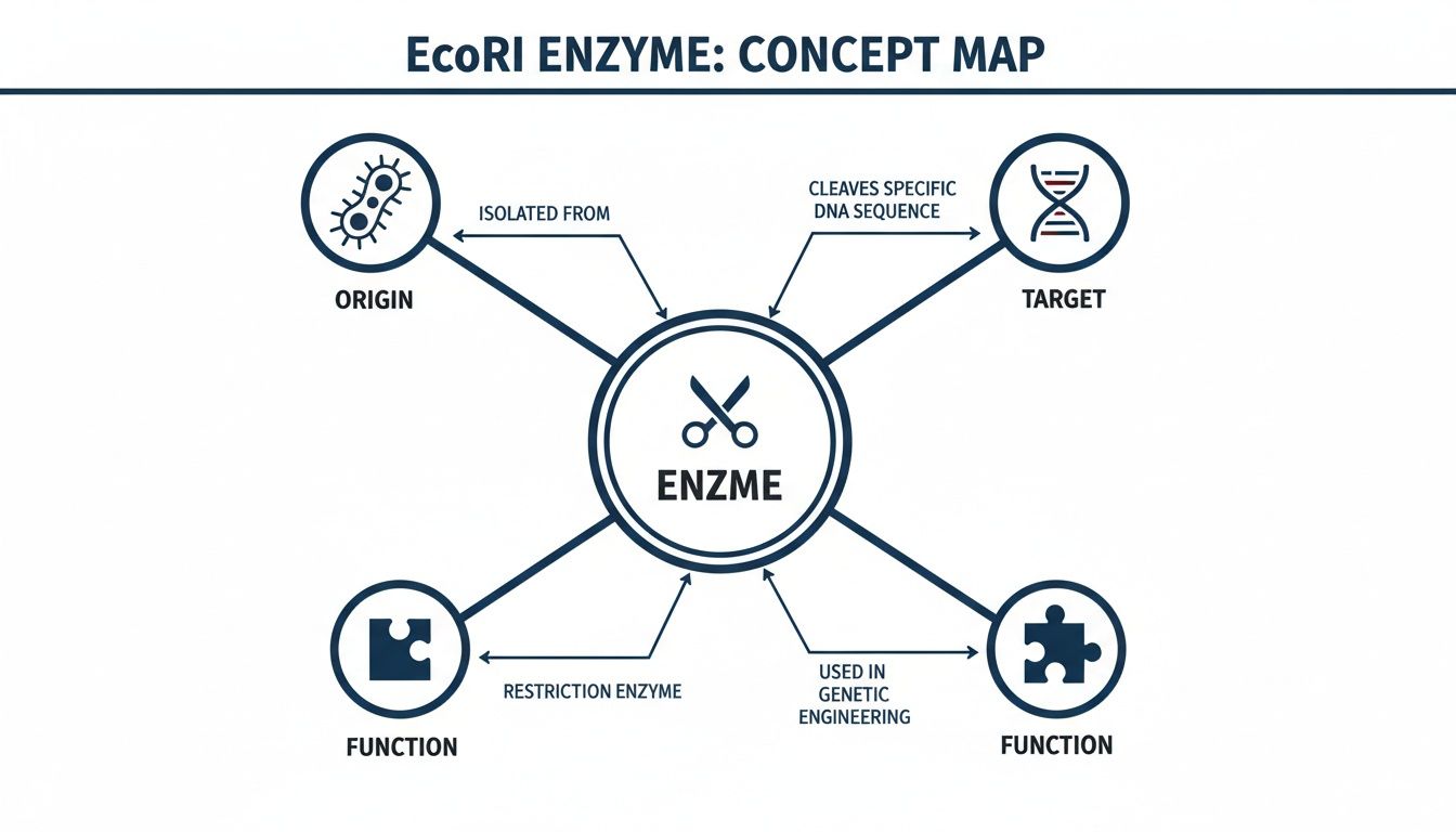
As you can see, the enzyme from E. coli acts like molecular scissors on DNA, creating the puzzle-piece function essential for genetic engineering.
The Power of Directional Cloning
The ability to create these predictable, complementary ends is what makes EcoRI so indispensable for gene cloning. When you use the same enzyme to cut both your gene of interest and your target plasmid vector, you guarantee they have matching sticky ends. This simple step dramatically increases the efficiency and accuracy of ligation—the process of "pasting" the gene into the plasmid.
This directional control is essential for creating functional expression vectors. By ensuring the gene is inserted in the correct orientation, you can guarantee that the cellular machinery will read the genetic code properly to produce the desired protein in your mammalian cell system.
Without these defined sticky ends, your gene could ligate in backward, leading to a completely non-functional protein and a failed experiment. The precise AATT overhangs generated by EcoRI provide a reliable, time-tested mechanism for building genetic constructs with confidence, a fundamental process that underpins countless applications in molecular biology.
Setting Up Your EcoRI Restriction Digest
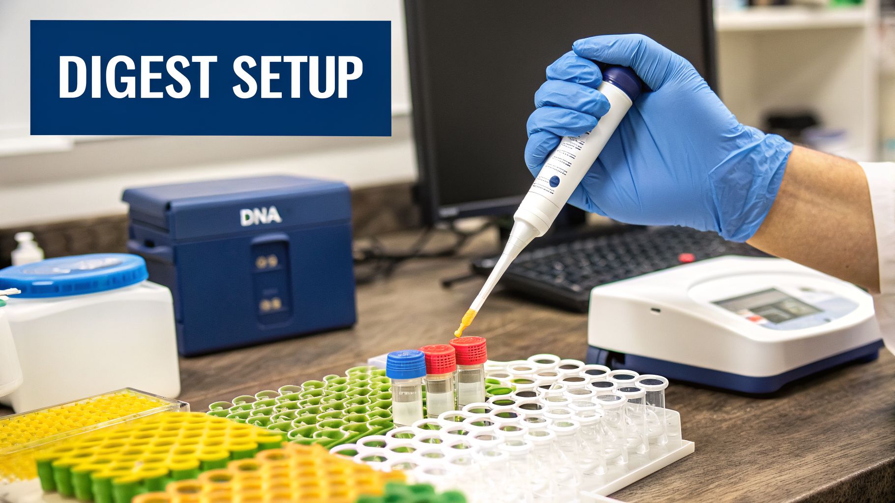
Theory is one thing, but getting a clean, complete digest on the bench is what really matters. A successful EcoRI restriction enzyme experiment comes down to a well-planned protocol where every component is respected. Think of it as a precise recipe—the right ingredients, in the right order, are the difference between a perfect cut and a failed experiment.
The setup itself is simple. You just need your DNA template, the enzyme, its specific reaction buffer, and some nuclease-free water to hit your final volume. But getting the balance and handling of these few components right is absolutely critical for a clean, complete cut.
The Essential Ingredients for Digestion
First up is your DNA template. This could be a plasmid you’re prepping for cloning, a PCR product you need to check, or genomic DNA for an RFLP. The single most important factor here is purity. Contaminants left over from your DNA purification—like salts, ethanol, or detergents—can easily inhibit or completely kill your enzyme’s activity.
Next is the star of the show: the EcoRI restriction enzyme. These enzymes are precious. They arrive highly concentrated and are suspended in glycerol to keep them stable at -20°C. It is absolutely crucial to keep the enzyme tube on ice at all times when it's out of the freezer. Always add it to your reaction mix last to prevent it from losing activity or starting the reaction before everything is ready.
Finally, there’s the reaction buffer, the unsung hero of the digest. This solution creates the perfect chemical environment—the right pH and salt concentration—for EcoRI to do its job efficiently. Most suppliers, like PurMa Biologics, will provide a 10X concentrated buffer with their enzyme, which you’ll dilute to 1X in your final reaction.
What Are Enzyme Units?
When you check the label on your vial of EcoRI, you'll see its concentration measured in "units/µL." This term can seem a bit abstract, but it's actually a very practical definition.
One unit of a restriction enzyme is defined as the amount of enzyme needed to completely digest 1 microgram (µg) of a specific substrate DNA (usually lambda phage DNA) in one hour at its optimal temperature.
For most routine plasmid digests, a good rule of thumb is to use 10–20 units of enzyme for every 1 µg of DNA. This usually works out to just 1 µL of enzyme. Using a slight excess like this gives you a safety margin, ensuring the digest goes to completion even if your DNA concentration isn't perfect. With high-quality modern enzymes, you don't really need to worry about over-digestion, but dumping in a massive excess is just a waste of an expensive reagent.
A Standard EcoRI Digestion Protocol
Let's put all of this into a practical, everyday protocol. Here’s a standard recipe for a 20 µL analytical digest of a plasmid. You can easily scale these volumes up or down for your specific needs, just be sure to maintain the same proportions.
Standard EcoRI Digestion Protocol Setup
| Component | Volume / Amount | Notes |
|---|---|---|
| Nuclease-Free Water | To 20 µL | Add water first, then buffer and DNA. Add the enzyme last. |
| 10X Reaction Buffer | 2 µL | Provides the optimal environment for enzyme activity. |
| DNA Template | 1 µg (e.g., 2 µL of 500 ng/µL) | Ensure DNA is high quality with a 260/280 ratio of ~1.8. |
| EcoRI Restriction Enzyme | 1 µL (10–20 units) | Keep on ice and add last. Mix gently by pipetting or flicking. |
After you've combined all the components, give the tube a gentle mix by flicking it with your finger or pipetting up and down a few times. Then, give it a quick pulse in a microcentrifuge to collect all the liquid at the bottom of the tube.
Incubate the reaction at 37°C for one hour. Once the hour is up, you can stop the reaction by adding a loading dye and putting the tube on ice, or you can kill the enzyme for good by heat-inactivating it at 65°C for 20 minutes before running your gel or moving on to a cleanup step.
How to Prevent EcoRI Star Activity
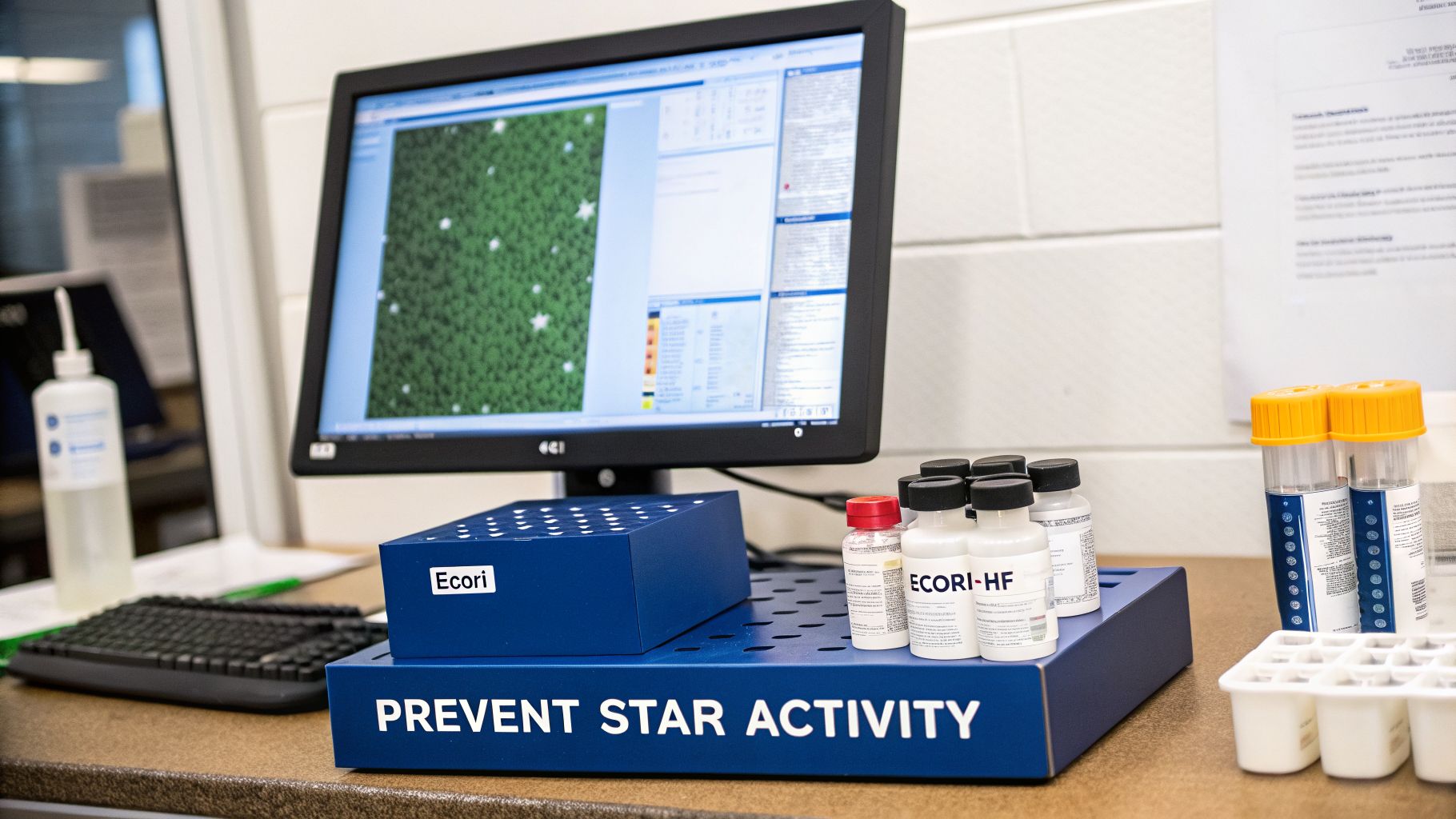
While the EcoRI restriction enzyme is famous for its precision, it has a notorious dark side: star activity. This is what happens when the enzyme gets sloppy, losing its strict specificity and cutting DNA at sites that are almost right but not quite its G'AATTC target.
Suddenly, your surgical scalpel starts behaving like a pair of dull scissors. It creates unexpected DNA fragments that can completely derail a cloning experiment or generate false positives in a diagnostic assay.
This off-target cutting isn't random. It’s a direct result of putting the enzyme in suboptimal, "uncomfortable" reaction conditions. Think of it like a key that, if you jiggle it with enough force, can sometimes open a lock it wasn't perfectly designed for. This relaxation of specificity is a major headache for any scientist counting on clean, predictable DNA cuts.
Common Triggers for Star Activity
Several factors in your reaction setup can push a standard EcoRI restriction enzyme into star activity. Knowing these triggers is the first step to avoiding the problem altogether. The usual suspects all relate to disrupting the carefully balanced chemical environment of the digest.
Key culprits include:
- High Glycerol Concentration: EcoRI is stored in a buffer with 50% glycerol to keep it from freezing solid at -20°C. If your enzyme volume makes up more than 10% of the total reaction, the final glycerol concentration can creep above 5%, a primary trigger for star activity.
- Incorrect Buffer or pH: Using a buffer with the wrong salt concentration or a pH outside the optimal 7.5–8.5 range can destabilize the enzyme, causing it to lose fidelity.
- High Enzyme-to-DNA Ratio: It’s tempting to dump in extra enzyme to make sure a digest goes to completion, but this can backfire. Too many enzyme molecules looking for a limited number of true sites increases the odds that some will bind and cut near-miss sequences.
- Prolonged Incubation Time: A one-hour digest is standard. Leaving a reaction with too much enzyme to run for hours or overnight gives it more time to make those undesirable off-target cuts.
The Modern Solution: High-Fidelity Enzymes
For decades, good lab practice meant meticulously controlling all these factors to keep star activity at bay. The modern solution, however, is far more direct and reliable: just use an engineered High-Fidelity (HF) version of the enzyme.
These HF enzymes are the result of targeted protein engineering. Scientists introduced specific mutations that essentially "sharpen" the enzyme's proofreading and recognition abilities.
Think of an HF enzyme as a finely-tuned instrument. It's been modified at the protein level to be more rigid and demanding about the sequence it recognizes. This makes it far less likely to lower its standards, even under conditions that would cause the original wild-type enzyme to start making mistakes.
The impact of the original EcoRI restriction enzyme on biotechnology was massive, creating a multi-billion dollar industry that reshaped mammalian cell research and biomanufacturing. Given its importance—and the persistent challenge of star activity—researchers spent over 30 years refining it.
They cloned and sequenced genes to develop high-fidelity versions that specifically address EcoRI's notorious tendency to cleave similar sites, like N'AATTC. Under bad conditions, star activity can affect 10-20% of cuts made by the wild-type enzyme. Modern HF mutants have virtually eliminated this problem, making them indispensable for reproducible science. You can read more about the development of these advanced enzymes.
Today, suppliers like PurMa Biologics typically offer these superior HF versions at the same price as the old wild-type enzyme. This makes the choice a no-brainer.
By opting for an EcoRI-HF restriction enzyme, you're proactively designing failure out of your experiment. It adds a crucial layer of security, ensuring your results are clean and free from the artifacts of star activity—something that's non-negotiable in regulated environments where accuracy is paramount. For virtually every application today, from routine plasmid checks to complex cloning projects, the HF version should be your default.
How EcoRI Is Used in Gene Cloning and Diagnostics
Understanding the mechanics of an EcoRI restriction enzyme is one thing, but seeing it in action is where you grasp its true power. This single enzyme is the backbone of countless molecular biology workflows, from building custom genes from scratch to diagnosing inherited diseases. For nearly fifty years, its precision and reliability have made it a workhorse in labs everywhere.
Its most common application, without a doubt, is gene cloning. Let's say your goal is to produce a human protein—like a monoclonal antibody—in mammalian cells. First, you need a way to get the gene that codes for that antibody into a circular piece of DNA called a plasmid vector, which will then carry your genetic payload into the cells.
This is exactly where the EcoRI restriction enzyme comes in. In a classic cloning workflow, you use EcoRI to cut both your gene of interest and the plasmid vector. Since both pieces of DNA are cut with the same enzyme, they now have perfectly complementary "sticky ends"—that signature 5'-AATT overhang. This shared sequence dramatically increases the odds that your gene will insert itself into the plasmid in the correct orientation.
Building Expression Vectors for Mammalian Systems
Once your gene is cut and ready, it can be ligated, or "pasted," into the opened-up plasmid. This step creates a brand-new, engineered molecule: a recombinant plasmid, also known as an expression vector. This vector is now a functional genetic blueprint, designed to do one specific job: tell a cell to start making your protein of interest.
This fundamental technique underpins some of the most advanced work in biotechnology today:
- Custom Protein Expression: Scientists build mammalian expression vectors to produce nearly any protein imaginable for research, diagnostics, or therapeutic development.
- Monoclonal Antibody Production: The process is absolutely essential for creating the hybridoma cell lines that generate highly specific antibodies used to treat cancer and autoimmune diseases.
- Stable Cell Line Generation: By introducing these vectors into mammalian cells, researchers can create stable cell lines that continuously produce a target protein—a critical step for any large-scale biomanufacturing operation.
The biochemical precision of EcoRI is why it's considered a 'workhorse' of molecular biology. Detailed kinetic studies show it cleaves both DNA strands at a rate of 0.35 s⁻¹ in specific lab assays, ensuring high specificity even on very long DNA strands. For teams in biomanufacturing, this translates to faster, more reliable gene cloning in mammalian systems. You can learn more about the enzyme kinetics that drive these applications.
The ability to build these vectors with such high fidelity is precisely why the EcoRI restriction enzyme is so important for labs and companies providing cell culture services. The high-quality vectors it helps create are the perfect starting point for boosting protein yields, often by 20–50%, when combined with advanced transfection reagents and specialized media in purification workflows.
A Powerful Tool in Genetic Diagnostics
Beyond cloning, EcoRI plays a vital role in diagnostic techniques—most notably Restriction Fragment Length Polymorphism (RFLP). This method cleverly uses the enzyme's precise cutting ability to spot variations in DNA sequences between different people.
The process itself is beautifully simple. A person's DNA is isolated and then digested with EcoRI. If a genetic mutation happens to have altered a G'AATTC recognition site, the enzyme can no longer cut there. As a result, the DNA fragments produced will be a different length compared to the fragments from a person without that mutation.
When you separate these fragments by size using gel electrophoresis, the difference becomes obvious as a distinct banding pattern. This allows scientists and clinicians to identify genetic markers linked to inherited diseases, run paternity tests, and even conduct forensic analysis. RFLP analysis using EcoRI turns a simple DNA cut into a powerful diagnostic indicator, connecting basic molecular biology directly to clinical applications.
Troubleshooting Your EcoRI Digestion
We’ve all been there. You set up your digest, run the gel, and your heart sinks. A single, stubborn band of uncut plasmid stares back at you. Or maybe you see a confusing mix of faint, correct bands alongside a bright band of starting material. Or worse, the dreaded smear.
When your EcoRI restriction enzyme digest doesn't work, it feels like your project has hit a brick wall. But before you throw out the enzyme, let's walk through a systematic checklist to figure out exactly what went wrong and get you back on track.
The first clue is always on your gel. Are you seeing zero cutting at all? Or just a partial, incomplete reaction? Each scenario points to a very different set of culprits.
Diagnosing Incomplete or No Digestion
If your EcoRI restriction enzyme isn't cutting — or is only giving you a weak, partial digest — it’s almost always one of a few usual suspects. Let’s work through them, starting with the most common offenders.
- Dead Enzyme: Enzymes are finicky proteins. If it was left on the bench too long, stored at the wrong temperature, or freeze-thawed one too many times, your EcoRI might simply be inactive.
- Dirty DNA: This is the big one. Your plasmid prep might be contaminated with leftovers from the purification kit. Things like ethanol, salts, or chaotropic agents are notorious enzyme inhibitors. A low 260/230 ratio from your NanoDrop is a major red flag.
- No Cut Site: It sounds simple, but it happens more than you'd think. Always run your sequence through a program like SnapGene or Benchling to confirm the G'AATTC site is actually there. It’s entirely possible the site was mutated out, or was never in your construct to begin with.
Before you ever blame the enzyme, interrogate your DNA. A quick sanity check of your DNA purity (aim for a 260/280 of ~1.8 and a 260/230 above 2.0) can save you from repeating a failed experiment with the exact same bad template.
If you suspect your DNA prep is the problem, here's a quick diagnostic test. Set up a small side reaction using a control plasmid you know cuts cleanly with EcoRI. If that control plasmid also fails to cut when you spike in a bit of your experimental DNA, you’ve just confirmed your prep has an inhibitor. The only real solution is to go back and re-purify your DNA.
Solving Smears and Unexpected Bands
What if your gel looks even weirder? A blurry smear down the lane or a ladder of bands you can't explain almost always points to one of two issues: nuclease contamination or star activity.
A smear is the classic signature of nucleases chewing up your DNA indiscriminately. This contamination can come from anywhere—your water, your buffer, even your pipette tips. This is why it's non-negotiable to use certified nuclease-free water and reagents for all your molecular biology work.
The other possibility is the classic sign of star activity. If you got a little heavy-handed with the enzyme, used the wrong buffer, or let the glycerol concentration get too high (above 5%), your EcoRI restriction enzyme may have started cutting at sites that look a bit like its real target. This off-target cutting creates a mess of unexpected bands.
The fix here is two-fold: first, double-check your reaction math and setup. Second, and more reliably, switch to a High-Fidelity (HF) version of the enzyme. These are engineered to be far less prone to star activity, even under slightly imperfect conditions. By working through these problems one by one, you’ll pinpoint the source of your trouble and get back to seeing those crisp, perfect bands you need.
Frequently Asked Questions About EcoRI
Even when you know the theory, using the EcoRI restriction enzyme on the bench can throw you a curveball. Here are some straight answers to the most common questions that pop up in the lab, designed to help you get cleaner, more reliable digests.
Can I Use a Universal Buffer for My EcoRI Digest?
Yes, you absolutely can. Modern universal buffers, like NEB's rCutSmart™ Buffer, are designed to give you 100% activity for EcoRI and hundreds of other enzymes. It’s what makes setting up a double digest so much easier, saving you from the headache of sequential reactions or dealing with compromise buffers.
That said, it's always good lab practice to quickly check the manufacturer's spec sheet for your specific enzyme and buffer lot. It’s a two-second check that confirms you’re getting the optimal performance you expect.
Why Is My DNA Not Cutting With EcoRI?
A failed digest is frustrating, but there are usually only a few culprits. The trick is to work through them systematically to figure out what went wrong.
First, the simple stuff: use your sequence analysis software to confirm there’s actually a G'AATTC site in your plasmid. It sounds obvious, but it’s a mistake that happens more often than you’d think and can save you a lot of troubleshooting.
Second, look at your DNA purity. Contaminants left over from your plasmid prep—like ethanol or excess salt—are notorious enzyme inhibitors.
Aim for a 260/280 ratio of ~1.8, but pay even closer attention to your 260/230 ratio. It should be well above 2.0. A low 260/230 is the classic sign of salt or solvent contamination that will kill your restriction digest.
Third, question your enzyme's activity. Did it get left out on the bench too long? Has it gone through too many freeze-thaw cycles? Improper storage is a fast way to inactivate it. Finally, while it’s rare for an EcoRI restriction enzyme, double-check if any methylation on your DNA could be blocking that specific recognition site.
What Is the Difference Between Standard and EcoRI-HF?
EcoRI-HF (High-Fidelity) is the engineered, next-generation version of the enzyme. It was specifically created to eliminate "star activity"—the tendency of the original, wild-type enzyme to make off-target cuts at the wrong sites when reaction conditions aren't perfect. Those non-specific cuts can completely ruin a cloning experiment.
The "HF" version has specific mutations that make it much more specific and forgiving, even if your reaction has a bit too much glycerol or runs longer than planned. It just gives you cleaner results and far more reproducible experiments.
Since most major suppliers now offer the HF version at the same price as the old standard enzyme, it has become the gold standard. For almost any application you can think of, EcoRI-HF is the smart choice to ensure your cuts are exactly where you want them.
How Should I Store My EcoRI Enzyme?
Proper storage is non-negotiable if you want your enzyme to work reliably. The EcoRI restriction enzyme must be stored in a non-frost-free freezer at -20°C at all times.
The glycerol in the storage buffer keeps it from freezing solid, which is why you can pipette it right out of the freezer. When you need it, take the tube out, place it immediately in a benchtop cooler (a "cold block"), and put it right back in the freezer the second you’re done. Never let it sit at room temperature, not even for a few minutes.
At PurMa Biologics, we know that reproducible science starts with reliable reagents. Our portfolio of high-quality buffers, molecular-grade water, and other cell culture essentials is built to support your research from discovery to production. We focus on ensuring you get clean, consistent results every single time. Explore our solutions to see how we can help you streamline your lab's workflows.