

How to Eradicate Mycoplasma in Your Lab: A 5-Step Protocol From 30 Years of Expertise
Mycoplasma contamination isn't something you can fix once and forget. It requires a systematic, ongoing approach — one that addresses incubator hygiene, incoming cell lines, routine screening, personal protective practices, and surface decontamination as a unified protocol. Here's how to implement it.
✅ Key Takeaways
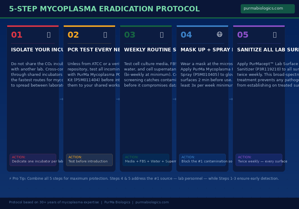
Isolate your incubator: Never share a CO₂ incubator with another lab. Cross-laboratory sharing is one of the fastest routes for mycoplasma to enter your cultures — a single contaminated flask from a neighboring lab can infect everything inside.
Test before you trust: Every cell line entering your lab from an external source must be quarantined and tested with a PCR-based mycoplasma PCR detection and elimination kit before introduction to shared equipment.
Screen weekly, not reactively: Your cell culture media, premium fetal bovine serum, water, and cell supernatants should all be tested for mycoplasma at least every two weeks — weekly is the recommended standard.
You are the biggest risk: Over 80% of lab personnel carry mycoplasma. Wearing a mask at the microscope and decontaminating gloves and surfaces with PurMa Mycoplasma Removal Spray before every session directly addresses the primary contamination source.
Sanitize everything, twice weekly: Applying PurMacept™ Lab Surface Sanitizer to all lab surfaces creates a hostile environment for any pathogen — providing a broad-spectrum second layer of defense beyond targeted mycoplasma removal.
Prevention beats treatment: Eradicating mycoplasma from contaminated live cultures is possible but laborious. Building a prevention-first protocol is faster, cheaper, and far more reliable.
Why You Need a Systematic Protocol — Not Just Good Intentions
Every cell culture lab has some awareness of mycoplasma contamination. Most researchers have encountered it at some point. Many have lost weeks of work to it. Yet despite this widespread awareness, contamination rates remain stubbornly high — with published studies consistently reporting that 15–35% of continuous cell cultures are contaminated at any given time. In some surveys, the number exceeds 60%.
The reason for this gap between awareness and action is straightforward: knowing that mycoplasma is a problem is not the same as having a protocol to prevent it. Good intentions — “we should test more often,” “we should be more careful” — don’t translate into contamination-free labs. What works is a defined, repeatable set of practices that every person in the lab follows, every day, without exception.
The protocol outlined in this article is built on three decades of hands-on mycoplasma research and product development at PurMa Biologics. It addresses the five primary vectors through which mycoplasma enters and spreads in mammalian cell culture laboratories, and for each vector, it provides a specific, actionable countermeasure. The steps are ordered from infrastructure-level prevention (incubator isolation) through to daily personal practices (masking and surface decontamination), creating a layered defense that is far more effective than any single measure alone.
Step 1: Isolate Your CO₂ Incubator — Do Not Share With Another Lab
This is the most impactful infrastructure decision you can make, and it’s the one most often compromised in shared laboratory facilities. The CO₂ incubator is not just a warm box — it’s an enclosed ecosystem with internal fans, air currents, and humidity that actively circulate particles between every flask, plate, and dish stored inside.
When you share an incubator with another lab, you are placing the integrity of your entire cell culture program in someone else’s hands. If a researcher from a neighboring lab introduces a contaminated flask — even unknowingly — the incubator’s internal air circulation will distribute mycoplasma-containing particles throughout the chamber. Research has demonstrated that airborne particles and aerosols generated during culture manipulations are among the greatest sources of microbial contamination, and incubator fans actively amplify this effect every time the door opens and closes.
The recommendation is absolute: dedicate one CO₂ incubator per laboratory. If complete separation isn’t immediately feasible, at minimum ensure that cultures from different labs are isolated in sealed containers within the incubator — never open plates or unsealed dishes alongside another lab’s cultures. But understand that sealed containers are a compromise, not a solution. True isolation is the only reliable prevention.
Even labs with excellent aseptic technique can be contaminated through a shared incubator. You can control your own practices perfectly and still have mycoplasma introduced by another group that doesn’t test as rigorously. Incubator isolation protects you from risks you cannot control.
Additionally, implement a regular incubator cleaning schedule. Remove all vessels, shelves, and trays periodically, and decontaminate internal surfaces with proven disinfectants. PurMa Biologics manufactures a proprietary CO₂ incubator treatment specifically designed to maintain pathogen-free conditions inside the chamber — a critical complement to the physical isolation strategy.
Step 2: PCR Test Every Incoming Cell Line Before It Enters Your Lab
The second most common route for mycoplasma to enter your lab isn’t through the air or from your skin — it’s through the front door, carried in a flask of cells from another laboratory. Published literature consistently identifies previously contaminated cell cultures from other research groups as the single largest external source of mycoplasma contamination. A single infected line, once introduced, can compromise every other culture in the facility within weeks.
Cell lines obtained from reputable repositories — such as ATCC, DSMZ, or other established biobanks — undergo rigorous mycoplasma testing before distribution and can generally be trusted. However, the reality of research is that many valuable and irreplaceable cell lines come from collaborator labs, core facilities, or legacy freezer stocks with uncertain provenance. These lines must be treated as potentially contaminated until proven otherwise.
PurMa Mycoplasma PCR Detection Kit
Cat# P5M011404
This kit provides species-level identification of mycoplasma contamination — not just a positive/negative result. Knowing whether you’re dealing with M. orale (human origin, likely from lab personnel) or M. arginini (bovine origin, likely from serum) tells you where the contamination came from and how to prevent recurrence. PCR-based detection delivers results within hours, versus the 4–5 weeks required by traditional agar culture methods.
The protocol is simple: every cell line from an external source gets quarantined in a separate incubator or sealed container and tested with the PurMa Mycoplasma PCR Detection Kit before it is allowed anywhere near your working cultures. No exceptions. It doesn’t matter how trusted the collaborator is, how reputable the institution is, or how urgently you need the cells. Test first. Introduce second. This single practice has prevented more contamination events than any other measure in cell culture history.
Step 3: Implement Weekly Routine Screening of All Reagents and Cultures
Testing incoming cell lines is essential, but it’s not sufficient. Mycoplasma can enter your cultures at any time — through contaminated reagents, through human transmission during routine handling, through cross-contamination from shared equipment. The only way to catch contamination before it compromises your data is through consistent, scheduled screening.
The recommended testing schedule is weekly. At an absolute minimum, screen every two weeks. Your testing should cover four critical targets:
Cell culture media — Test each batch of cell culture media, whether commercially purchased or prepared in-house using proprietary chemically defined media formulations. Contamination can be introduced during handling, aliquoting, or storage.
Fetal bovine serum (FBS) — Premium fetal bovine serum is a known potential vector for bovine-origin mycoplasma species, particularly M. arginini and A. laidlawii. Even reputable serum suppliers cannot guarantee 100% mycoplasma-free product without independent verification on your end.
Cell culture water — Water used in media preparation, washing, and other cell culture applications should be tested regularly. Contaminated water sources can introduce mycoplasma that persists through subsequent media preparation steps.
Cell supernatants — This is your most direct indicator. Testing the supernatant of your actively growing cultures tells you whether the cells themselves — the cultures producing your experimental data — are currently contaminated.
The most effective approach is to designate a specific day each week as “mycoplasma testing day” and make it non-negotiable. Monday mornings work well — it catches any contamination that may have been introduced during the previous week while giving you time to respond before critical experiments later in the week. Document every test result. A contamination log is both a quality control tool and an early warning system that reveals trends before they become crises.
Step 4: Mask Up and Decontaminate Surfaces Before Every Session
This step addresses the single greatest source of mycoplasma contamination in any lab: you.
The data is unambiguous. Over 80% of laboratory technicians carry mycoplasma in their oral cavity and oropharynx. The most commonly isolated species — M. orale, M. fermentans, and M. salivarium — are human commensals that colonize healthy individuals without causing symptoms. You don’t know you’re carrying them. You can’t prevent yourself from carrying them. But you can prevent them from reaching your cultures.
Wear a mask when checking cells under the microscope. This is the single most exposed moment in cell culture work. You’re leaning directly over your cultures, often for extended periods, breathing directly onto flask surfaces and the surrounding bench area. A mask — even a standard surgical mask — creates a physical barrier that dramatically reduces the transfer of oral mycoplasma to your workspace.
But masking alone isn’t enough. Mycoplasma that has already been deposited on surfaces can remain viable for four to six days, waiting to be transferred to your cultures via gloves, pipettors, or any other point of contact. Surface decontamination before each work session is essential.
PurMa Mycoplasma Removal Spray (Mycospray)
Cat# P5M010405
Apply to gloves, hood surfaces, microscope stages, pipettors, and all working surfaces at minimum 2 minutes before use. PurMa Mycoplasma Removal Spray is formulated to destroy mycoplasma at the DNA level, achieving 100% elimination of A. laidlawii, M. arginini, M. bovis, M. fermentans, M. hominis, M. hyorhinis, and M. orale. Unlike standard 70% ethanol, which has limited and unreliable efficacy against mycoplasma, this formulation specifically targets the organisms you’re most concerned about. It’s safe for skin contact and produces no harmful vapors at room temperature. Use at least three times per week.
Apply PurMa Mycoplasma Removal Spray to all surfaces and let it sit for a full 2 minutes before beginning work. Do not wipe it dry — let the formulation work. This brief waiting period is the difference between surface disinfection and merely wetting the surface. Build it into your routine: spray, set a timer, prepare your materials, and begin work when the timer sounds.
Step 5: Apply Broad-Spectrum Lab Surface Sanitization Twice Weekly
Steps 1 through 4 target mycoplasma specifically. Step 5 broadens the defense to cover all pathogens — because mycoplasma is not the only contaminant threatening your cultures, and a truly clean lab requires comprehensive surface management.
PurMacept™ Lab Surface Sanitizer (10X)
Cat# P3R119210
This proprietary lab surface sanitizer is designed for twice-weekly application to all lab surfaces — benchtops, incubator exteriors, equipment surfaces, door handles, refrigerator handles, and any other shared contact points. Unlike the targeted mycoplasma action of PurMa Mycoplasma Removal Spray, PurMacept™ provides broad-spectrum pathogen elimination that prevents any microorganism from establishing on treated surfaces. The two products are complementary: Mycospray for immediate pre-session decontamination, PurMacept™ for ongoing environmental control.
The logic behind twice-weekly application is straightforward. Mycoplasma can survive on surfaces for up to six days. By sanitizing every three to four days, you ensure that no surface in your lab remains untreated long enough for deposited organisms to accumulate and pose a contamination risk. This creates an environment where even if mycoplasma is introduced — through a visitor, a new technician, or a momentary lapse in masking protocol — it cannot persist long enough to reach your cultures.
Think of it this way: Steps 1–3 prevent mycoplasma from entering and detect it early. Step 4 blocks the primary transmission pathway. Step 5 ensures that whatever gets through the first four layers doesn’t survive to cause contamination. It’s defense in depth — and it works.
When Contamination Is Found: Triage and Treatment
Even with the best prevention protocol, contamination can occasionally occur. The critical factor isn’t whether it ever happens — it’s how quickly you detect it and how decisively you respond.
First Response: Quarantine Immediately
The moment a culture tests positive for mycoplasma, remove it from the shared incubator and isolate it completely. Do not wait for a confirmatory test. Do not continue working with it “just for this one experiment.” Mycoplasma spreads through aerosols, surface contact, and shared equipment — every hour a contaminated culture remains in your workspace increases the risk of cross-contamination to everything around it.
Decision Point: Discard or Treat?
For most contaminated cultures, the safest and most efficient response is to discard the affected cells and restart from clean frozen stocks. If you’ve followed good cell banking practices — maintaining seed stocks from verified clean sources — this allows rapid recovery with minimal disruption.
However, there are situations where disposal isn’t an option: stable transfectants developed over months, irreplaceable hybridomas, monoclonal antibody-producing lines, or primary cultures that cannot be re-derived. For these high-value lines, specialized treatment is justified.
The PurMa Mycoplasma Treatment Kit (MTK, 100X) provides a sequential three-phase treatment protocol — MTR-I, followed by MTR-II, then MTR-III — designed to eliminate mycoplasma from live cells without destroying the host culture. The treatment is effective across multiple mycoplasma species, but it requires careful adherence to the protocol and post-treatment retesting to confirm successful eradication. As PurMa Biologics emphasizes from three decades of working with this pathogen: treatment is possible, but prevention is always preferable.
Don’t just treat the affected culture — treat the environment. Perform a thorough decontamination of the incubator, hood, and all shared equipment using PurMa Mycoplasma Removal Spray. Apply PurMacept™ Lab Surface Sanitizer to all lab surfaces. Then test your remaining cultures to assess whether the contamination has spread. A contamination event is both a problem to solve and an opportunity to reinforce your prevention protocol.
The Complete Ecosystem: Why Reagent Quality Matters
Mycoplasma prevention doesn’t exist in isolation from the rest of your cell culture practice. The quality and integrity of every reagent you use — from cell culture media and premium fetal bovine serum to cell culture suited buffers, cell culture suited PBS, and cell culture suited antibiotics — contributes to the overall resilience of your cultures against contamination.
Poorly manufactured reagents can introduce contaminants directly. Inconsistent reagent quality can stress cells, making them more susceptible to mycoplasma-induced damage. And reagents prepared without adequate quality control may contain trace endotoxins, heavy metals, or other impurities that compromise cell health in ways that mimic — or compound — the effects of mycoplasma contamination.
This is why PurMa Biologics approaches cell culture as a complete ecosystem rather than a collection of individual products. From proprietary chemically defined media manufactured under stringent quality control, to water and buffers tested for endotoxin and mycoplasma contamination, to the proprietary CO₂ incubator treatment that keeps your incubator environment pathogen-free — every element is designed to work together to maintain the conditions under which healthy, uncontaminated cells produce reliable, reproducible data.
“The fight against mycoplasma isn’t won by a single product or a single practice. It’s won by a system — a layered, comprehensive approach where every element of your cell culture workflow contributes to contamination prevention. That’s what three decades of working with this pathogen has taught us.”
When your mammalian cell culture reagents are manufactured to the highest standards, your detection protocols are rigorous, your surfaces are decontaminated, and your personal practices are disciplined, mycoplasma contamination becomes manageable — not inevitable. That shift in mindset, from reactive to preventive, is ultimately what separates labs that struggle with contamination from labs that don’t.
Frequently Asked Questions
CO₂ incubators with internal fans and air circulation actively spread mycoplasma-containing particles every time the door opens. A single contaminated flask from another lab can aerosolize mycoplasma throughout the entire chamber, contaminating every culture inside — including yours. Dedicated incubators eliminate this cross-laboratory transmission pathway entirely. If sharing is unavoidable, use only sealed flasks (never open plates) and isolate your cultures in a closed plastic container within the incubator.
Extremely quickly. Research has shown that after trypsinizing a single contaminated culture in a laminar flow hood, live mycoplasma could be recovered from the flask exterior, hemocytometer, pipettor, pipette discard pan, and the hood surface — where they survived for four to six days. A clean culture subcultured just once per week in the same hood tested positive for mycoplasma after only six weeks. This demonstrates how a single contaminated flask can compromise an entire lab in a matter of weeks.
Standard 70% ethanol is a general disinfectant effective against many bacteria and viruses, but its efficacy against mycoplasma is limited and inconsistent. Because mycoplasma lack a cell wall — which is the primary target for many disinfectants — they respond differently than typical bacteria. Purpose-built mycoplasma decontamination sprays like PurMa Mycoplasma Removal Spray are specifically formulated to destroy the DNA composition of all major mycoplasma species with verified 100% efficacy. For reliable mycoplasma elimination on surfaces, ethanol alone is not sufficient.
Test four critical targets: cell culture media, fetal bovine serum (FBS), cell culture water, and cell supernatants from actively growing cultures. The recommended frequency is weekly, with bi-weekly as the absolute minimum. Additionally, every new cell line from any external source must be quarantined and tested before introduction to shared incubators and hoods. Designating a specific day each week as “mycoplasma testing day” helps ensure consistency.
Over 80% of laboratory technicians carry mycoplasma — primarily M. orale — in their oral cavity and oropharynx. When you lean over cultures at the microscope, your breath introduces these organisms directly onto flask surfaces and the surrounding workspace. A mask creates a physical barrier that blocks this primary contamination pathway. It’s the simplest, least expensive, and most immediately effective protective measure available.
These are complementary products designed for different purposes. PurMa Mycoplasma Removal Spray (Mycospray, Cat# P5M010405) is specifically formulated to destroy mycoplasma DNA and should be applied to gloves, hood surfaces, microscope areas, and immediate workspaces 2 minutes before each work session, at least 3 times per week. PurMacept™ Lab Surface Sanitizer (Cat# P3R119210) is a broad-spectrum solution that eliminates all pathogens from any treated surface, applied twice weekly to benchtops, equipment, and all shared contact points. Together, they create a two-layer defense: targeted mycoplasma elimination for active work sessions, plus ongoing broad-spectrum environmental protection.
The safest first response is always to discard contaminated cultures and restart from clean frozen stocks. This eliminates the contamination definitively and avoids the risk of incomplete treatment. However, for irreplaceable lines — stable transfectants, hybridomas, monoclonal antibody producers — the PurMa Mycoplasma Treatment Kit (MTK, 100X) provides a sequential three-phase treatment (MTR-I → MTR-II → MTR-III) that can eliminate mycoplasma from live cells. Post-treatment, cultures must be passaged without antibiotics and retested to confirm successful eradication before reintroduction to the general lab environment.
References
-
Nikfarjam L, Farzaneh P. Prevention and Detection of Mycoplasma Contamination in Cell Culture. Cell Journal (Yakhteh). 2012;13(4):203–212.
https://pmc.ncbi.nlm.nih.gov/articles/PMC3584481/ -
Huang X, et al. Prevention, Diagnosis and Eradication of Mycoplasma Contamination in Cell Culture. Journal of Biological Methods. 2023.
https://pmc.ncbi.nlm.nih.gov/articles/PMC10668599/ -
Olarerin-George AO, Hogenesch JB. Assessing the prevalence of mycoplasma contamination in cell culture via a survey of NCBI's RNA-seq archive. Nucleic Acids Research. 2015;43(5):2535–2542.
https://academic.oup.com/nar/article/43/5/2535/2453278 -
ATCC. Mycoplasma Contamination — Detection, Prevention, and Elimination.
https://www.atcc.org/the-science/authentication/mycoplasma-contamination -
Uphoff CC, Drexler HG. Eradication of Mycoplasma Contaminations from Cell Cultures. Current Protocols in Molecular Biology. 2014;106:28.5.1–28.5.12.
https://www.dkfz.de/genomics-proteomics/genomics-proteomics/fileadmin/ccontrol/user_upload/Eradication_of_Mycoplasma.pdf -
Uphoff CC, Drexler HG. Cell Culture Mycoplasmas. Deutsche Sammlung von Mikroorganismen und Zellkulturen (DSMZ).
https://www.dsmz.de/fileadmin/user_upload/Collection_MuTZ/CellCulture_Mycoplasma.pdf - Rottem S, Barile MF. Beware of Mycoplasmas. Trends in Biotechnology. 1993;11:143–151.
-
PurMa Biologics. Mycoplasma Detection and Elimination Reagents.
https://purmabiologics.com/mycoplasma-detection-and-elimination/ -
Hay RJ. Contamination of Tissue Cultures by Mycoplasmas. In: Contamination of Cell Cultures. IntechOpen; 2012.
https://www.intechopen.com/chapters/40228