

Mycoplasma in Cell Culture: The Silent Pathogen Destroying Your Research From Within
It doesn't cloud your media. It doesn't kill your cells overnight. It doesn't trip a single alarm. And that's exactly what makes mycoplasma the most devastating contaminant in mammalian cell culture today.
📋 Key Takeaways
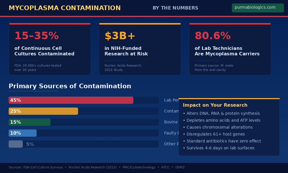
Invisible threat: Mycoplasma contamination affects 15–35% of continuous cell cultures worldwide, yet produces no visible signs in culture media — making it the most under-detected pathogen in research labs.
Your results are compromised: Contaminated cultures produce wildly inconsistent experimental readouts. What you may attribute to faulty cell culture media or premium fetal bovine serum could be mycoplasma quietly degrading your cells.
You are the source: Over 80% of lab technicians carry mycoplasma in their oral cavity. Human breath — specifically M. orale and M. fermentans — accounts for the majority of contamination events.
Standard antibiotics don’t work: Mycoplasma lack cell walls, rendering penicillin and most common cell culture suited antibiotics completely ineffective against them.
Detection is everything: PCR-based mycoplasma PCR detection and elimination reagents are the gold standard for identifying contamination — and should be part of your weekly lab routine.
PurMa Biologics brings three decades of hands-on expertise in manufacturing the most reliable mycoplasma detection, treatment, and surface elimination products available today.
What Exactly Is Mycoplasma — and Why Should You Care?
If you work with mammalian cell culture, there’s a reasonable chance that right now — as you read this — mycoplasma is actively growing inside your incubator. Not maybe. Not hypothetically. The published data suggests that between 15% and 35% of continuous cell cultures worldwide are contaminated at any given time. In some regional surveys, that number has climbed past 60%.
Mycoplasma are the smallest self-replicating organisms known to science. With over 200 identified species, these parasitic bacteria belong to the class Mollicutes, a name that literally translates to “soft skin” — because they lack a rigid cell wall entirely. This single biological trait is what makes them so insidious in the context of cell culture: they slip through standard filtration membranes (at just 0.3–0.8 µm in diameter), they resist the antibiotics your lab already uses, and they produce absolutely no turbidity in your cell culture media.
Think about that for a moment. A pathogen that can be present at concentrations 1,000 times greater than your actual cells — and you would never see it looking at your flask.
The Silent Destroyer: Why Mycoplasma Is So Dangerous
Bacterial contamination? You see the cloudiness within hours. Fungal contamination? Those hyphae are hard to miss. But mycoplasma operates on a completely different playbook. It’s a pathogen that has evolved to survive inside its host’s environment without triggering the obvious warning signs that other contaminants produce. And that’s precisely what makes it the most destructive organism you will ever encounter in cell culture work.
The damage mycoplasma inflicts is cumulative and multi-dimensional. At the cellular level, contamination leads to altered DNA, RNA, and protein synthesis pathways. It diminishes amino acid availability and depletes ATP levels — effectively starving your cells of the energy they need to function normally. It causes chromosomal alterations that fundamentally change the biological behavior of your culture. And because mycoplasma depend on host cells for survival (they lack the genes for synthesizing their own macromolecule precursors), they actively hijack your cells’ metabolic machinery to sustain themselves.
The result? A cell culture that looks passable on the surface but is biologically compromised at every level. Your cells are not performing as they should. Your data is not reliable. And the worst part is: you have no idea.
The Incubation Trap: It’s Growing While You’re Not Looking
Here’s the scenario that plays out in labs every single day: a researcher receivefs a new cell line, perhaps from a collaborator’s lab, and introduces it into the shared CO₂ incubator. Everything looks fine. Cells are adhering, proliferating, and passaging normally. Two weeks pass. Then four. Then six. Slowly, imperceptibly, something starts to change.
Mycoplasma has one of the longest effective incubation periods of any cell culture contaminant. Unlike bacteria that announce their presence within 24–48 hours through media turbidity, mycoplasma can establish itself in your culture over weeks or even months before any detectable effect becomes apparent. During this entire period, it’s replicating, competing for nutrients in your cell culture media and premium fetal bovine serum, and subtly reprogramming your cells’ gene expression.
By the time you notice something is “off” with your cultures — slower growth, unusual morphology, inconsistent assay results — the contamination may have been present for weeks. Every experiment you ran during that period is potentially compromised. Every dataset, every conclusion, every publication.
This long incubation window is precisely why reactive approaches to mycoplasma don’t work. You cannot wait until you suspect contamination to start testing. By then, the damage is done — and it has almost certainly spread to other cultures sharing the same incubator, hood, and workspace.
The Great Misdiagnosis: When You Blame the Media, Not the Microbe
This is perhaps the most costly misconception in cell culture today, and it happens far more frequently than anyone wants to admit: a researcher notices their cells aren’t growing well. Viability is dropping. Passage times are extending. The immediate, reflexive conclusion? “The media must be bad.” Or: “This new batch of FBS isn’t working.” Or: “The supplier changed something.”
In reality, the cell culture media, the premium fetal bovine serum, the cell culture suited buffer, and the cell culture suited PBS may all be performing exactly as specified. The true culprit — mycoplasma — is quietly degrading your cells from within, and its effects mimic the symptoms of poor reagent quality almost perfectly.
Consider the parallels: mycoplasma competition for arginine depletes the media of this essential amino acid, leading to nutrient-deprived cells that grow poorly. Mycoplasma endonucleases degrade host cell DNA, creating the kind of erratic cellular behavior you’d associate with a bad lot of serum. Mycoplasma-induced cytokine disregulation can make cells behave as if the media formulation is off when, in fact, the formulation is fine — the cells are just responding to a pathogen you haven’t tested for.
“If your cells suddenly aren’t performing despite using the same reagents you’ve always used, the question shouldn’t be ‘what’s wrong with my media?’ The question should be: ‘when did I last test for mycoplasma?'”
This misdiagnosis creates a cascade of wasted resources. Labs discard perfectly good media. They switch suppliers. They reformulate protocols. They spend weeks troubleshooting a reagent problem that doesn’t exist — all while the actual contaminant continues to proliferate unchecked.
Fluctuating Results: The Hallmark of Hidden Contamination
If there’s one red flag that should immediately make you suspect mycoplasma, it’s this: your experimental readouts are inconsistent in a way that doesn’t track with any variable you can control.
Here’s what makes mycoplasma-contaminated data so treacherous. The pathogen doesn’t affect your cells uniformly or statically. As mycoplasma populations grow, plateau, and compete for resources within your culture, the degree of cellular disruption changes over time. An assay performed on Monday, when mycoplasma levels are at one stage of growth, may produce results dramatically different from the same assay performed on Thursday, when the pathogen population has shifted.
This creates a pattern of results that is the hallmark of mycoplasma contamination: high standard deviations, poor reproducibility between replicates, and readouts that fluctuate from experiment to experiment with no apparent cause. A 2015 study published in Nucleic Acids Research found that mycoplasma contamination significantly affected the expression of at least 61 host genes, with contaminated samples showing vastly different expression profiles compared to clean cultures. The financial implications are staggering — the same study estimated that over $3 billion in NIH-funded research could be affected by undetected mycoplasma contamination.
If your statistical analyses are showing unexpectedly high standard deviations, or if you’re seeing results that should be reproducible but aren’t, don’t just repeat the experiment. Test for mycoplasma first. It could save you weeks of troubleshooting — and rescue the integrity of your entire dataset.
The Human Factor: You Are the Primary Source
This is the part of the conversation that makes lab personnel uncomfortable, but it needs to be said directly: you are the single greatest source of mycoplasma contamination in your cell culture lab.
Research published in the journal Cytotechnology and compiled in PMC reviews has demonstrated that 80.6% of laboratory technicians are carriers of mycoplasma, primarily Mycoplasma orale — a species that colonizes the human oral cavity and oropharynx. M. orale consistently ranks as the leading contaminant in cell culture studies worldwide, accounting for 20–40% of all mycoplasma infections. Two other human species, M. fermentans and M. salivarium, are also frequently detected.
The transmission pathway is startlingly direct. Every time you lean over a culture flask to check it under the microscope, every time you speak while working at the hood, every time you handle materials without proper surface decontamination, you’re potentially introducing mycoplasma into your workspace. One study showed that after trypsinizing an infected culture, live mycoplasma could be recovered from the outside of the flask, the hemocytometer, the pipettor, the pipette discard pan, and the surface of the laminar flow hood — where they remained viable for four to six days.
Always wear a mask when checking cells under the microscope. This single practice can dramatically reduce the transfer of oral mycoplasma species from your respiratory tract to your cultures and work surfaces. It costs nothing, takes no extra time, and directly addresses the primary contamination pathway.
Once mycoplasma enters a single culture in your lab, the contamination spreads rapidly through shared equipment, shared hoods, and shared incubators. Within weeks, it’s possible for every culture in the lab to test positive for the same mycoplasma strain.
Why Standard Antibiotics Fail Against Mycoplasma
If you’re relying on your standard cell culture suited antibiotics — penicillin, streptomycin, and their common combinations — to protect against mycoplasma, you’re operating under a dangerous assumption. Here’s why:
Penicillin works by disrupting bacterial cell wall synthesis. Mycoplasma have no cell wall. The antibiotic has literally zero mechanism of action against them. Streptomycin shows activity against roughly half of mycoplasma strains but is completely ineffective against the others. Gentamicin, at the concentrations typically used in cell culture, is similarly unreliable. In practice, mycoplasma are resistant to most antibiotic cocktails routinely added to culture media.
This biological reality means that antibiotics give labs a false sense of security. Researchers who faithfully add penicillin/streptomycin to every flask may believe they’re protected, when in fact they’ve done nothing to address the one contaminant most likely to be present. Worse, the routine use of antibiotics can actually mask the symptoms of bacterial co-contamination that would otherwise serve as an early warning sign of broader aseptic technique problems in the lab.
Effective mycoplasma elimination requires specialized reagents designed to target organisms that lack cell walls — a very different pharmacological challenge than what standard antibiotics are designed to solve.
Detection Matters: The Case for Routine PCR Testing
Given everything we’ve discussed — the invisible growth, the gradual deterioration, the fluctuating results, the human transmission vector — the conclusion is inescapable: routine, proactive testing for mycoplasma is not optional. It is a fundamental requirement of responsible cell culture practice.
The gold standard for mycoplasma detection today is PCR-based testing. Unlike older methods such as direct culture on agar plates (which require 4–5 weeks of incubation) or fluorescent staining with Hoechst 33258 (which requires subjective visual interpretation), PCR provides rapid, sensitive, specific, and cost-effective detection that can identify contamination within hours rather than weeks.
The best PCR detection kits don’t just tell you whether mycoplasma is present — they identify the exact species responsible for the contamination. This matters because different species have different sources, and knowing which mycoplasma you’re dealing with can help you trace the contamination back to its origin and prevent recurrence.
Test your cell culture media, fetal bovine serum, water, and cell supernatants for mycoplasma at minimum every two weeks — and ideally weekly. Any new cell line entering the lab, regardless of its source, should be quarantined and tested before introduction to shared equipment. Cells obtained from repositories like ATCC are generally reliable, but lines received from other laboratories should always be treated as potentially contaminated until proven otherwise.
Three Decades of Expertise: How PurMa Biologics Leads the Fight
PurMa Biologics has spent over thirty years focused on a single mission: giving researchers the tools they need to detect, eliminate, and prevent mycoplasma contamination in cell culture. That depth of experience — three decades of working directly with this devastating pathogen — has produced a comprehensive line of mycoplasma PCR detection and elimination reagents that represent the current state of the art.
Detection: Know What You’re Dealing With
PurMa Mycoplasma PCR Detection Kit
Cat# P5M011404This sophisticated PCR-based kit doesn’t just detect the presence of mycoplasma — it identifies the exact species causing the contamination. Species-level identification is critical for tracing the source of infection and implementing targeted prevention strategies. Whether the source is human-origin (M. orale, M. fermentans), bovine-origin (M. arginini, A. laidlawii), or another vector entirely, this kit gives you the precise answer.
View Mycoplasma Detection & Elimination Reagents →Elimination: Remove Mycoplasma From Live Cultures
When contamination is confirmed and the affected cell line is too valuable to discard — stable transfectants, hybridomas, or other irreplaceable cultures — PurMa Biologics offers the PurMa Mycoplasma Treatment Kit (MTK) (100X), a three-phase treatment protocol (MTR-I → MTR-II → MTR-III) designed to eliminate mycoplasma from live cells without destroying the culture itself. The sequential treatment approach ensures thorough eradication across multiple mycoplasma species.
Surface Decontamination: Break the Transmission Cycle
PurMa Mycoplasma Removal Spray (Mycospray)
Cat# P5M010405Formulated to destroy mycoplasma at the DNA level, PurMa Mycospray eliminates A. laidlawii, M. arginini, M. bovis, M. fermentans, M. hominis, M. hyorhinis, and M. orale with 100% efficacy. Safe for skin contact and producing no harmful vapors at room temperature, it should be applied to gloves, hood surfaces, microscope areas, and all working surfaces at least 2 minutes before use. For maximum protection, use it at least three times per week.
Learn More About Mycoplasma Removal Spray →PurMacept™ Lab Surface Sanitizer (10X)
Cat# P3R119210For comprehensive, ongoing lab surface protection, PurMacept™ provides a proprietary lab surface sanitizer that eliminates not just mycoplasma but all pathogens from any treated surface. Applied twice weekly, it creates a hostile environment for microbial growth on benchtops, incubator surfaces, equipment, and any other shared lab surfaces. This is your second line of defense — a broad-spectrum surface treatment that complements the targeted mycoplasma action of Mycospray.
Learn More About Lab Surface Sanitizer →What sets PurMa Biologics apart isn’t just the breadth of the product line — it’s the depth of understanding behind it. Every product in the mycoplasma detection and elimination range has been developed by scientists who have spent careers working with this pathogen. That firsthand expertise translates into reagents that perform reliably under real-world lab conditions, not just under ideal test scenarios. With a complete ecosystem of mammalian cell culture reagents — from proprietary chemically defined media and premium fetal bovine serum to cell culture suited antibiotics, cell culture suited buffers, cell culture suited PBS, and proprietary CO₂ incubator treatment — PurMa Biologics provides everything needed to maintain clean, consistent, and reliable cell cultures.
Frequently Asked Questions
Studies consistently report that 15–35% of continuous cell cultures are contaminated with mycoplasma. The U.S. FDA tested over 20,000 cell cultures across three decades and found a 15% contamination rate. Regional surveys have reported rates as high as 60–80%, particularly in labs without routine testing protocols. Even a conservative estimate of 11%, derived from an unbiased NCBI RNA-seq archive survey, confirms that mycoplasma contamination remains a pervasive problem in modern research.
Mycoplasma cells are extremely small — just 0.3 to 0.8 µm in diameter — placing them below the resolution limit of standard light microscopy. They also lack a rigid cell wall, making them pleomorphic (irregularly shaped) and difficult to distinguish from cellular debris. Crucially, mycoplasma do not cause turbidity in culture media, meaning there is no visible change to alert you. PCR-based detection kits are the only reliable method for routine mycoplasma screening.
Standard cell culture antibiotics like penicillin target bacterial cell wall synthesis — and since mycoplasma lack a cell wall entirely, penicillin has no effect. Streptomycin inhibits roughly half of mycoplasma strains but fails against the rest. Gentamicin is generally ineffective at concentrations used in routine cell culture. Effective mycoplasma elimination requires specialized treatment reagents, such as the PurMa Mycoplasma Treatment Kit (MTK), that target these wall-less organisms through alternative mechanisms.
At minimum, test every two weeks. Weekly testing is ideal and strongly recommended for labs performing sensitive assays or maintaining valuable cell lines. Your testing should cover cell culture media, FBS, cell culture water, and cell supernatants. Additionally, every new cell line entering the lab — regardless of source — should be quarantined and tested before being introduced to shared equipment or incubators.
Laboratory personnel are the primary source. Published research demonstrates that over 80% of lab technicians carry mycoplasma, particularly M. orale, in their oral cavity. Human breath during routine lab activities — checking cells under the microscope, speaking at the hood — introduces these organisms directly into the workspace. Once on a surface, mycoplasma can remain viable for four to six days, providing ample opportunity for cross-contamination.
Profoundly. Mycoplasma contamination alters DNA, RNA, and protein synthesis; depletes amino acids and ATP; causes chromosomal changes; and can disregulate hundreds of host genes. This produces dramatically inconsistent experimental readouts with high standard deviations — results that fluctuate based on the stage of mycoplasma growth rather than your experimental variables. A single study estimated that over $3 billion in NIH-funded research could be impacted by undetected mycoplasma contamination.
Reputable repositories like ATCC maintain rigorous quality control and mycoplasma testing protocols, making them the safest source for cell lines. However, many valuable lines are only available through collaborator labs, and these carry significantly higher contamination risk. The practical rule: accept cell lines from any source you need, but always quarantine and test them with a reliable PCR detection kit — such as the PurMa Mycoplasma PCR Detection Kit — before introducing them to your lab’s shared incubator space.
References
-
Olarerin-George AO, Hogenesch JB. Assessing the prevalence of mycoplasma contamination in cell culture via a survey of NCBI's RNA-seq archive. Nucleic Acids Research. 2015;43(5):2535–2542.
https://academic.oup.com/nar/article/43/5/2535/2453278 -
Nikfarjam L, Farzaneh P. Prevention and Detection of Mycoplasma Contamination in Cell Culture. Cell Journal (Yakhteh). 2012;13(4):203–212.
https://pmc.ncbi.nlm.nih.gov/articles/PMC3584481/ -
Uphoff CC, Drexler HG. Cell Culture Mycoplasmas. Deutsche Sammlung von Mikroorganismen und Zellkulturen (DSMZ).
https://www.dsmz.de/fileadmin/user_upload/Collection_MuTZ/CellCulture_Mycoplasma.pdf -
ATCC. Mycoplasma Contamination — Detection, Prevention, and Elimination.
https://www.atcc.org/the-science/authentication/mycoplasma-contamination - Razin S, Kahane I, Banai M, Bredt W. Adhesion of Mycoplasmas to Eukaryotic Cells. Ciba Foundation Symposium. 1981;80:98–118.
-
PurMa Biologics. Mycoplasma Detection and Elimination Reagents.
https://purmabiologics.com/mycoplasma-detection-and-elimination/ - Rottem S, Barile MF. Beware of Mycoplasmas. Trends in Biotechnology. 1993;11:143–151.
-
Hay RJ. Contamination of Tissue Cultures by Mycoplasmas. In: Contamination of Cell Cultures. IntechOpen; 2012.
https://www.intechopen.com/chapters/40228